
Your heel bone absorbs up to 7 times your body weight with each step you take. That is a lot of repeated force on a single bone.
When that load builds up faster than the bone can repair itself, a calcaneal stress fracture forms.
The frustrating part? It often feels exactly like plantar fasciitis at first. Many people keep training on it, which turns a small crack into a serious injury.
This article breaks it all down: symptoms, causes, diagnosis, and what recovery actually looks like.
What Is a Calcaneal Stress Fracture?
A calcaneal stress fracture is a small crack inside the heel bone. It develops from repeated impact, not a single fall or accident.
The calcaneus is the largest bone in the foot. Every step sends force directly through it. Under normal conditions, the bone responds to this stress by rebuilding itself stronger.
The problem starts when activity increases too fast, or bone health is already compromised. Micro-damage builds up faster than the repair process can keep pace. A tiny crack forms in the heel bone and widens with each step.
Unlike a traumatic heel fracture from a fall, this injury develops gradually over days or weeks. Catching it early keeps recovery short. Missing it turns a six-week problem into a six-month one.
What Are the Symptoms of a Calcaneal Stress Fracture?
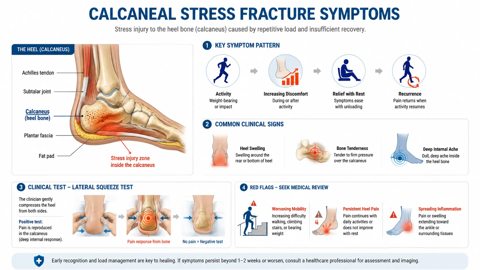
The most telling sign is a deep, aching pain in the heel that builds over the course of the day. It eases with rest. Then it returns the moment you load the foot again.
This pattern is different from most heel conditions. The pain does not hit hardest with your first steps in the morning. It gets progressively worse the longer you stay on your feet.
Other symptoms include:
- Swelling on the sides or bottom of the heel
- Tenderness when pressing directly on the heel bone
- A heavy, dull ache rather than a sharp, stabbing pain
- Pain at rest in more advanced cases
One clinical test stands out from the rest. A doctor applies pressure to both sides of the heel at the same time. Pain during this squeeze test is a strong indicator of a stress fracture. Plantar fasciitis rarely produces pain with this test. That condition affects the tissue along the bottom of the foot, not the sides of the bone.
When to See a Doctor:
Do not wait. Get checked promptly if you notice any of the following:
- Pain that does not improve after a full day off your feet
- Swelling that spreads toward the ankle
- Inability to put weight on the heel at all
- Symptoms that get worse after one to two weeks of rest
These signs suggest the fracture is progressing. Imaging is needed right away.
Calcaneal Stress Fracture vs. Plantar Fasciitis
Both conditions cause heel pain. Both can feel worse after a period of rest. That surface-level overlap is exactly what leads to missed diagnoses.
Misdiagnosing a stress fracture as plantar fasciitis keeps people active. That continued activity is what turns a manageable crack into a fracture requiring months of recovery.
The table below shows the key differences between the two conditions.
| Feature | Calcaneal Stress Fracture | Plantar Fasciitis |
|---|---|---|
| Pain location | Across the heel bone | Bottom of heel, near the arch |
| Pain pattern | Worsens through the day | Worst with first steps in the morning |
| Squeeze test | Often positive | Rarely positive |
| Pain at rest | Possible in advanced cases | Uncommon |
| Imaging needed | MRI or X-ray | Usually, clinical diagnosis only |
Achilles tendinopathy is another condition that gets confused with a calcaneal stress fracture. Achilles problems cause pain along the back of the heel and tendon, often with visible tendon swelling.
A stress fracture causes pain over the bone itself, not along a tendon. When the diagnosis is uncertain, imaging confirms which one it is.
What Causes a Calcaneal Stress Fracture?
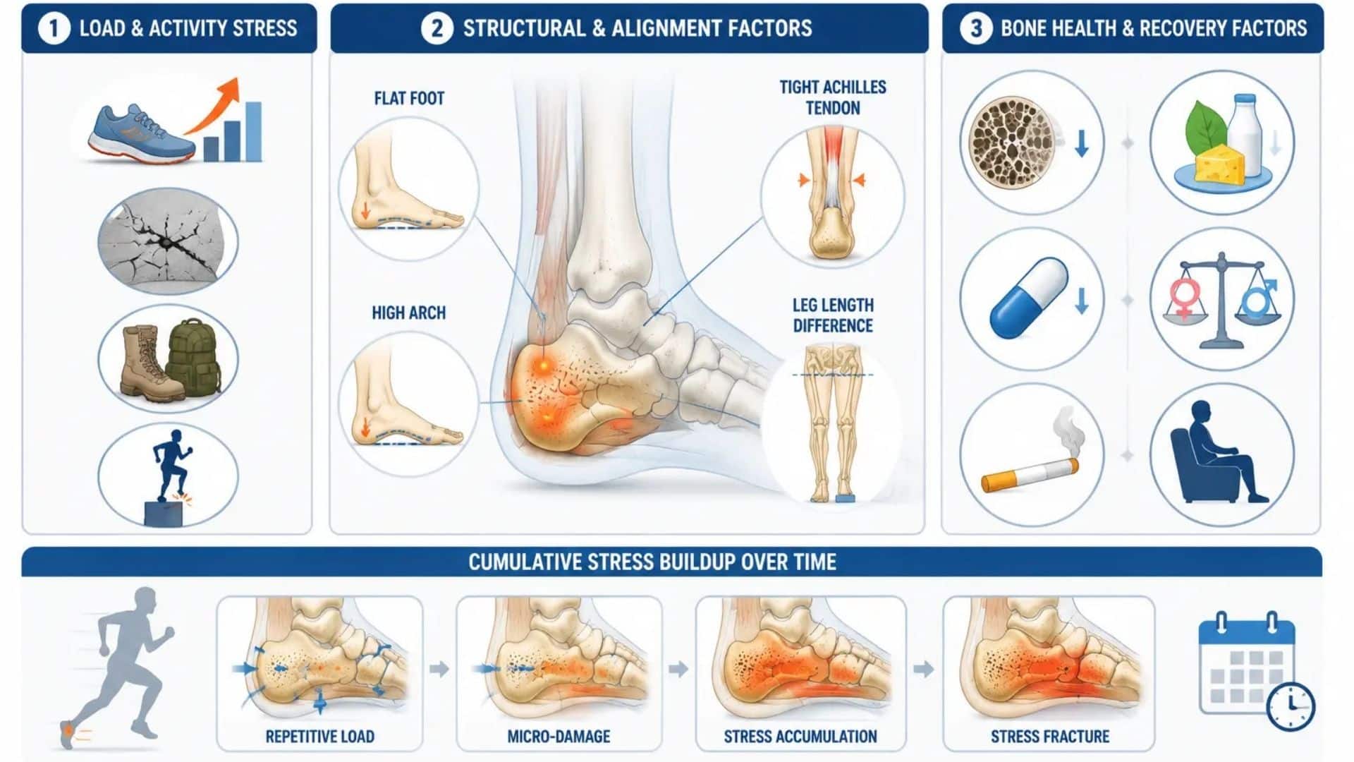
The root cause is always the same. The bone is asked to do more than it can currently handle, more often than it can recover from.
Training Mistakes That Raise Your Risk
Training errors are the most common trigger. These include:
- Increasing running mileage by more than 10 percent per week
- Starting a high-impact program without a gradual build-up
- Running long distances on hard surfaces like concrete
- Military training with heavy loads and long-distance marching
Biomechanical factors also contribute:
- Flat feet spread the load unevenly across the heel
- High arches concentrate impact directly through the calcaneus
- A tight Achilles tendon increases compression at the heel with each stride
- Leg length differences create imbalanced loading with every step
Bone Health and Medical Risk Factors
Some people carry a higher risk because of their bone health. Contributing factors include:
- Osteoporosis, which weakens bone structure even during low-intensity activity
- Low calcium and vitamin D intake, which slows the bone’s ability to repair micro-damage
- Long-term corticosteroid use, which reduces bone mineral density over time
- Hormonal changes, such as irregular periods in female athletes or low testosterone in males
- Low energy availability, which is a key concern in the Female Athlete Triad
Smoking and prolonged inactivity also reduce bone strength and increase overall fracture risk.
How Is a Calcaneal Stress Fracture Diagnosed?
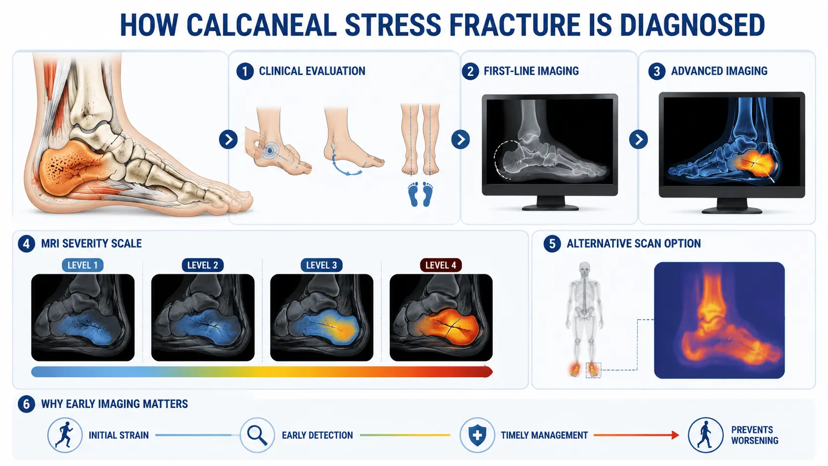
Diagnosing a calcaneal stress fracture involves a combination of physical examination and imaging tests, as early fractures are not always visible on standard X-rays.
1. Physical Examination
Your doctor checks for tenderness over the heel bone, performs the squeeze test, and looks for signs such as bruising on the sole (Mondor sign), swelling, limited movement, and changes in the way you walk.
2. Initial X-Ray
An X-ray is usually the first imaging test, but it often appears normal during the first two to four weeks after the injury. A visible fracture line or healing changes may only appear several weeks later.
3. MRI for Early and Accurate Diagnosis
If symptoms strongly suggest a stress fracture despite a normal X-ray, an MRI is the preferred test. It can detect bone stress within days of injury, confirm the diagnosis early, and determine the severity of the fracture.
4. MRI Grading
Doctors use MRI findings to classify the injury, which helps determine the most appropriate treatment plan.
| MRI Grade | What It Shows | What It Means |
|---|---|---|
| Grade 1 | Bone marrow swelling on one imaging sequence | Early stress reaction |
| Grade 2 | Swelling on multiple sequences without a fracture line | Moderate stress injury |
| Grade 3 | Visible fracture line with significant swelling | Established stress fracture |
| Grade 4 | Complete fracture line through the bone | A severe fracture that may require surgery |
5. Bone Scan When MRI Is Unavailable
A bone scan may be used if an MRI cannot be performed. It is sensitive for detecting increased bone activity but cannot confirm the exact cause as accurately as an MRI.
If heel pain continues despite a normal X-ray, further evaluation with an MRI can help confirm the diagnosis early and prevent the injury from progressing.
Calcaneal Stress Fracture Treatment
Most cases heal without surgery when treated early. Treatment focuses on protecting the heel, supporting bone healing, and returning to activity in a structured way.
- Rest and Activity Modification: Stop running and all high-impact activities immediately. Recovery typically takes two to eight weeks, depending on severity.
- CAM Walking Boot: A boot protects the heel during daily activities, reduces impact, and allows limited weight-bearing while the bone heals.
- Pain Management: Apply ice for 15 to 20 minutes several times per day. Use over-the-counter pain relievers only as directed by your doctor. Long-term NSAID use can affect bone healing, so check with your provider first.
- Nutritional Support: Get adequate amounts of calcium and vitamin D through food or supplementation, if needed. Nutritional deficiencies often contribute to this injury. Correcting them supports faster repair.
- Low-Impact Exercise: Swimming, cycling, and pool running are all safe options during recovery. These do not place stress on the heel. Stop any exercise that causes heel pain.
- Physical Therapy Rehabilitation improves strength, gait, and movement patterns. This reduces the chance of a repeat fracture after returning to training.
Surgery is rarely needed. It is reserved for severe or displaced fractures that cannot heal properly with conservative treatment.
Recovery Time and Return to Activity
Healing is confirmed by repeat imaging at six to eight weeks. Doctors look for new bone formation and reduced bone marrow swelling before clearing you to progress.
The table below shows what to expect at each phase of recovery.
| Phase | Timeframe | What to Do |
|---|---|---|
| Acute rest | Weeks 1 to 2 | Stop all impact. Use a boot or crutches as directed. |
| Protected weight-bearing | Weeks 2 to 6 | Walk-in boot. Begin pool running or cycling. |
| Transition | Weeks 6 to 8 | Confirm healing with imaging. Begin walking without boots. |
| Gradual return | Weeks 8 to 12 | Start a walk-to-run program. Increase by no more than 10 percent per week. |
| Full return | Week 12 onward | Return to full training. Watch for returning pain. |
Do not rush this. The fracture comes back easily if you return to full training before the bone is ready. If pain returns when activity increases, step back, rest for a few days, and rebuild more slowly.
How to Prevent a Calcaneal Stress Fracture
Train Gradually. Do not increase weekly mileage, session intensity, or training frequency by more than 10 percent in any given week. The bone needs time to adapt to new load demands.
Replace Your Footwear. Running shoes lose their shock absorption well before they look worn out. Replace them every 300 to 500 miles. Check with a specialist about [running shoe options](INTERNAL LINK 3) if you have flat feet or high arches. Orthotics can redistribute load more evenly across the foot.
Support Your Bone Health: Eat enough food. Get adequate calcium and vitamin D through diet or supplementation. Female athletes should watch for the warning signs of the Female Athlete Triad. These include low energy availability, irregular periods, and low bone density. The American College of Obstetricians and Gynecologists provides guidance on recognizing and addressing this condition.
Vary Your Training Surface. Hard surfaces like concrete increase impact on the heel with every stride. Mixing in softer surfaces, such as grass or running tracks, reduces the cumulative load on the bone.
Add Strength Training. Stronger foot and calf muscles absorb more force during each step. This reduces the share that goes directly to the bone. Two sessions per week of targeted lower-leg strength work make a measurable difference over time.
Conclusion
A calcaneal stress fracture is a serious overuse injury that is easy to underestimate in the early stages.
The heel bone handles more load per step than almost any other bone in the body. When it starts to crack, it needs time away from that load to repair itself.
Early diagnosis, proper imaging, a structured recovery, and a gradual return to activity are what separate a six-week setback from a six-month one.
If your heel has been aching for more than a week or two, especially if the pain worsens as the day goes on, get it checked.
Have you dealt with a calcaneal stress fracture? What helped most during your recovery? Share your experience in the comments below.
Frequently Asked Questions
Can a Calcaneal Stress Fracture Heal on Its Own?
Yes, with adequate rest and reduced activity, most cases heal without surgery. Continuing to load the heel without treatment is what causes a minor fracture to become a complete break.
How Long Does a Calcaneal Stress Fracture Take to Heal?
Most heal in six to eight weeks with proper rest and a protective boot. A full return to running typically takes 10 to 20 weeks, depending on the severity of the fracture.
Can I Exercise During Recovery?
Yes. Swimming, Cycling, and Pool Running Are All Safe Options as Long as They Do Not Cause Heel Pain. Staying Active During Recovery Makes the Return to Training Easier.
What Happens if A Calcaneal Stress Fracture Goes Untreated?
The fracture can progress to a complete break, causing severe pain, major swelling, and a possible need for surgery. Early treatment prevents the injury from reaching this stage.
What Is the Squeeze Test for A Calcaneal Stress Fracture?
A doctor compresses both sides of the heel at the same time. Pain during this test is a strong indicator of a stress fracture. Plantar fasciitis rarely produces pain with this maneuver.
Is a Calcaneal Stress Fracture the Same as Plantar Fasciitis?
No. A calcaneal stress fracture is a crack in the heel bone. Plantar fasciitis affects the connective tissue along the bottom of the foot. A squeeze test and imaging can confirm the correct diagnosis.